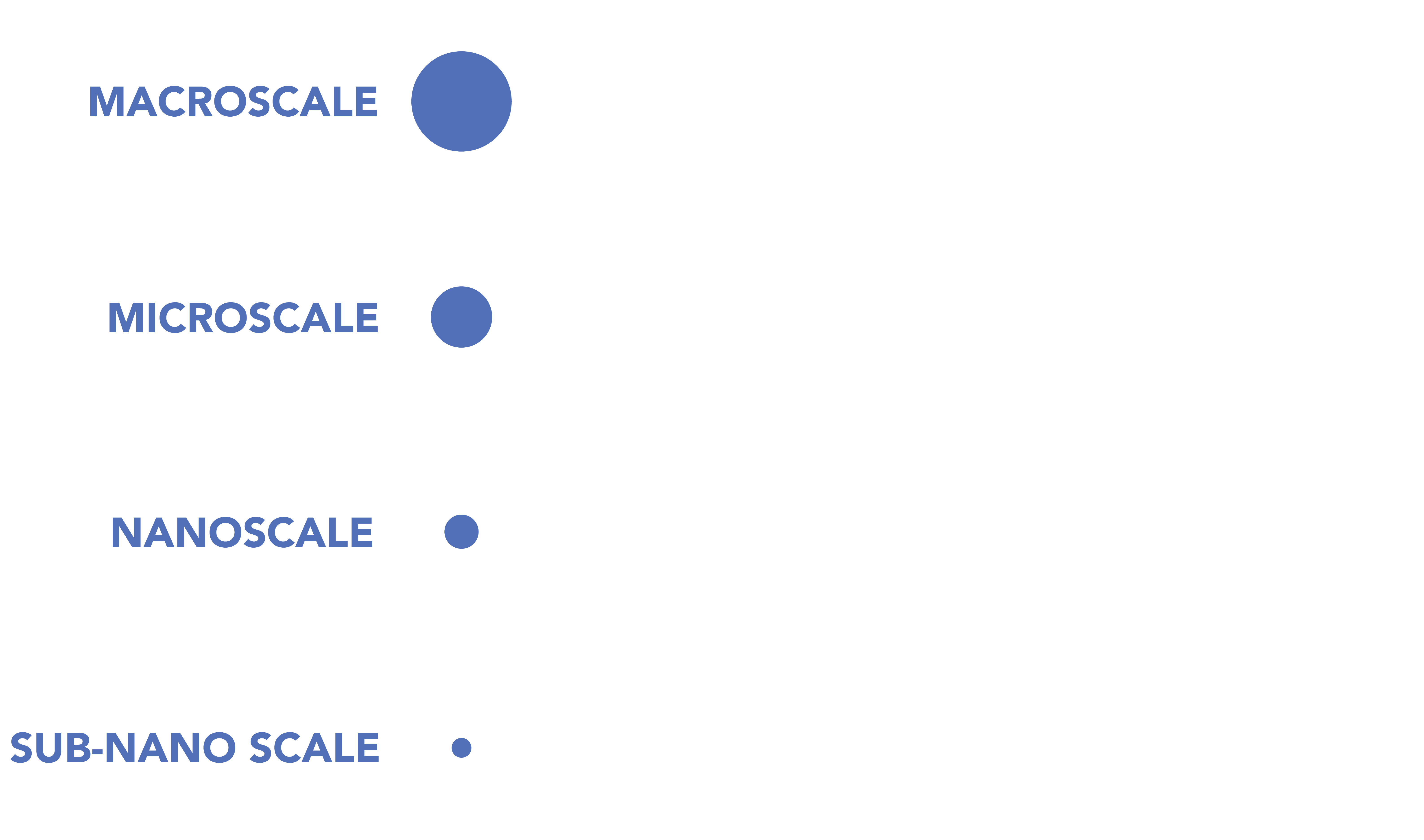
A multiscale, correlated imaging and spectroscopy suite to study biomaterials
from the macro to the nanoscale.
COCOON is a next-generation characterization suite designed for researchers working at the intersection of biology, materials science, and optics.
Built around a scanning electron microscope, COCOON integrates Raman spectroscopy, elemental analysis, and cryogenic sample preparation into a single unified platform.
This environment enables something rare: direct, real-time correlation of composition, structure, and function across orders of magnitude in scale, all within one workflow, at both room temperature and cryogenic conditions.
Researchers can move seamlessly from whole-sample imaging to nanoscale structural mapping, from molecular identity to elemental distribution, and from dynamic behavior to high-resolution visualization. This combination allows COCOON to reveal relationships in natural and engineered materials that would otherwise remain hidden.
COCOON is a Tufts University Core Facility, supported by the Office of Naval Research and the W. M. Keck Foundation.
COCOON enables a level of correlated multimodal analysis that is uncommon even in advanced microscopy centers. By tightly integrating optical, spectroscopic, elemental, and electron-based techniques, the platform allows researchers to:
Together, these tools deliver a comprehensive, multiscale picture of how organic, biological, and natural materials are built and how they function.
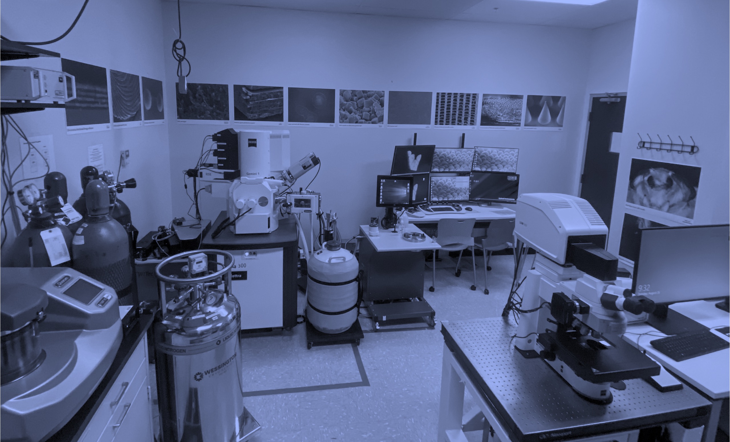
COCOON houses a tightly integrated set of instruments optimized for cross-correlated analysis. This integrated suite ensures that samples can be tracked across instruments without losing spatial context, enabling true correlative microscopy.
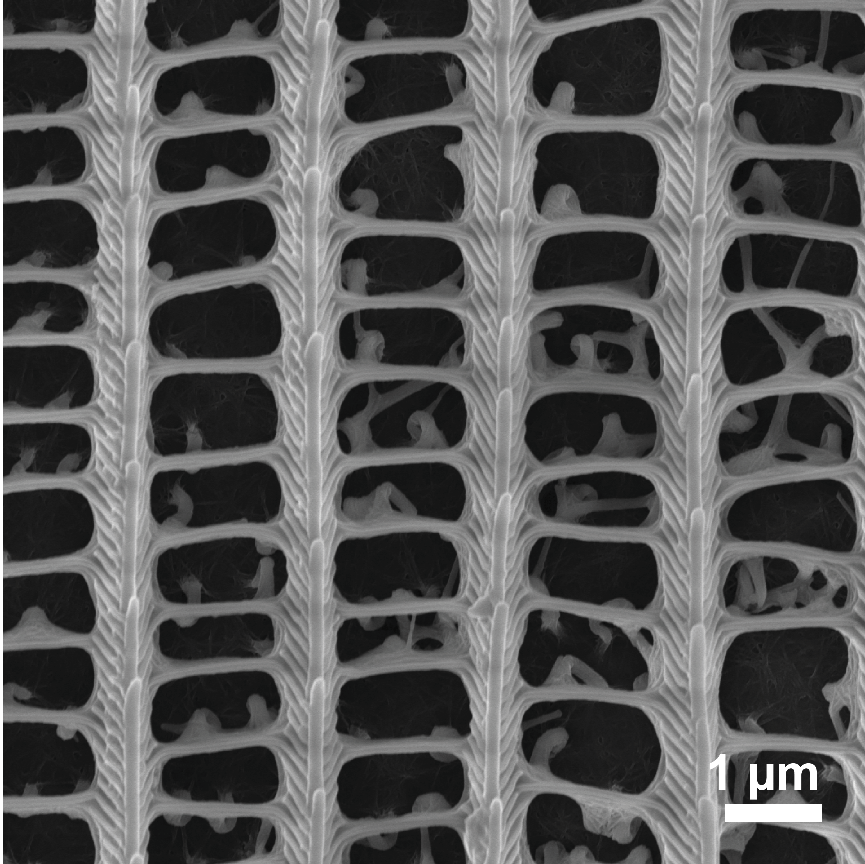
SEM image of silk aerogel
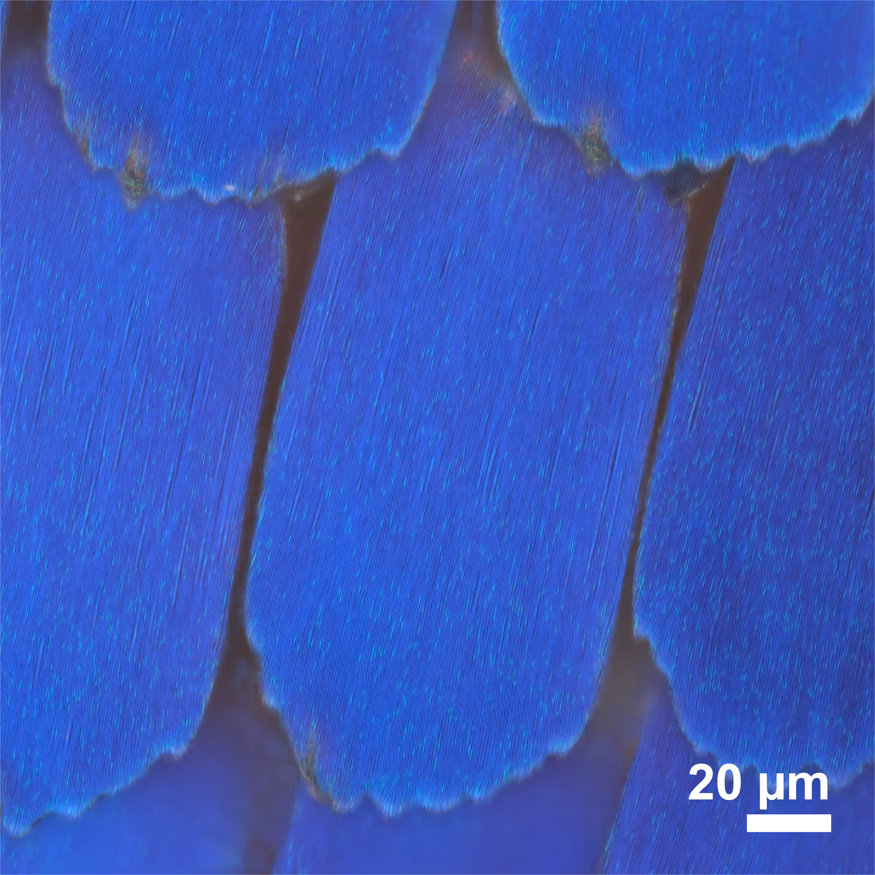
Bright-field reflection image of Morpho butterfly scales
SEM image of Morpho butterfly scale
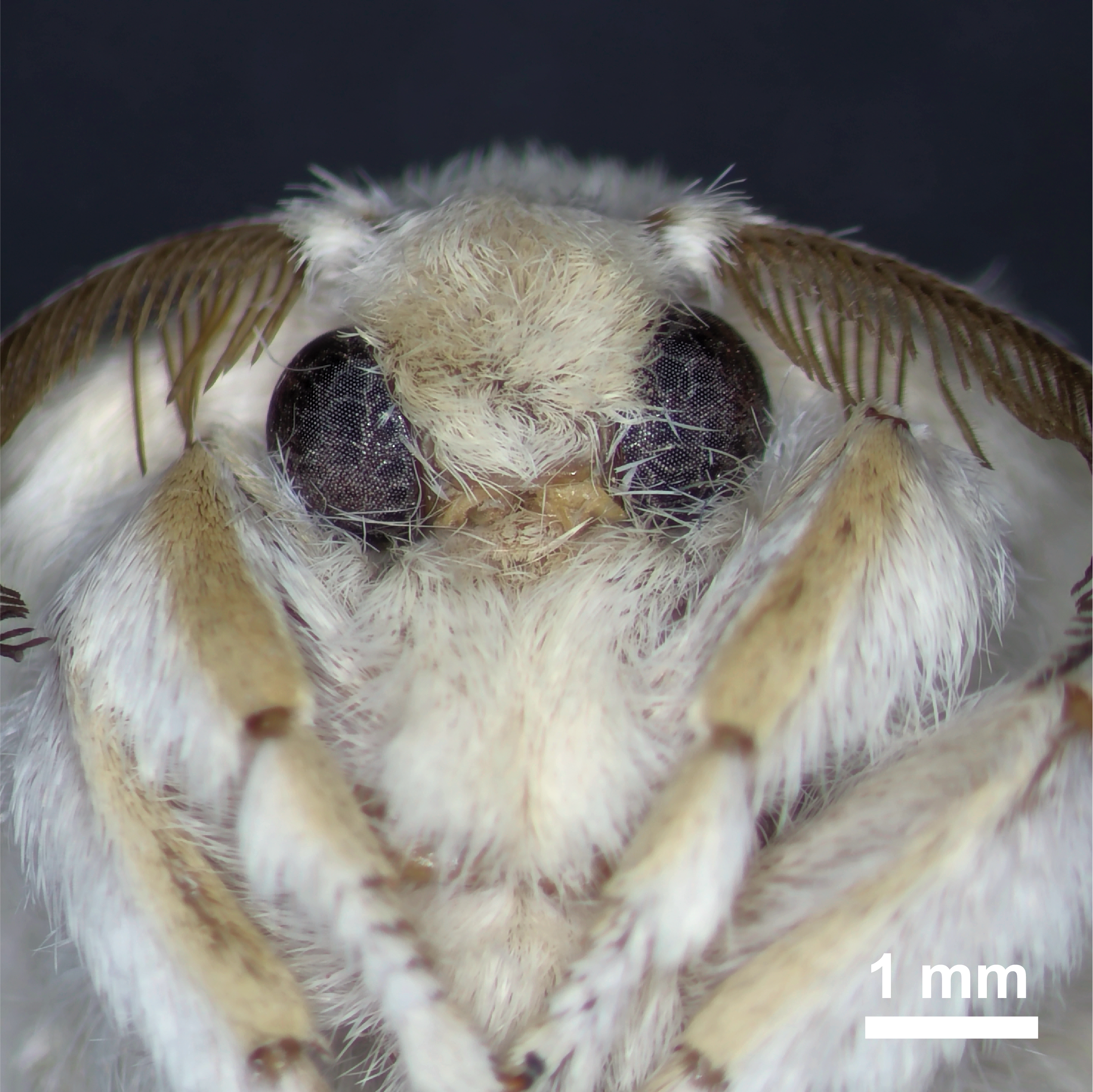
Stereomicroscopy image of Bombyx mori moth
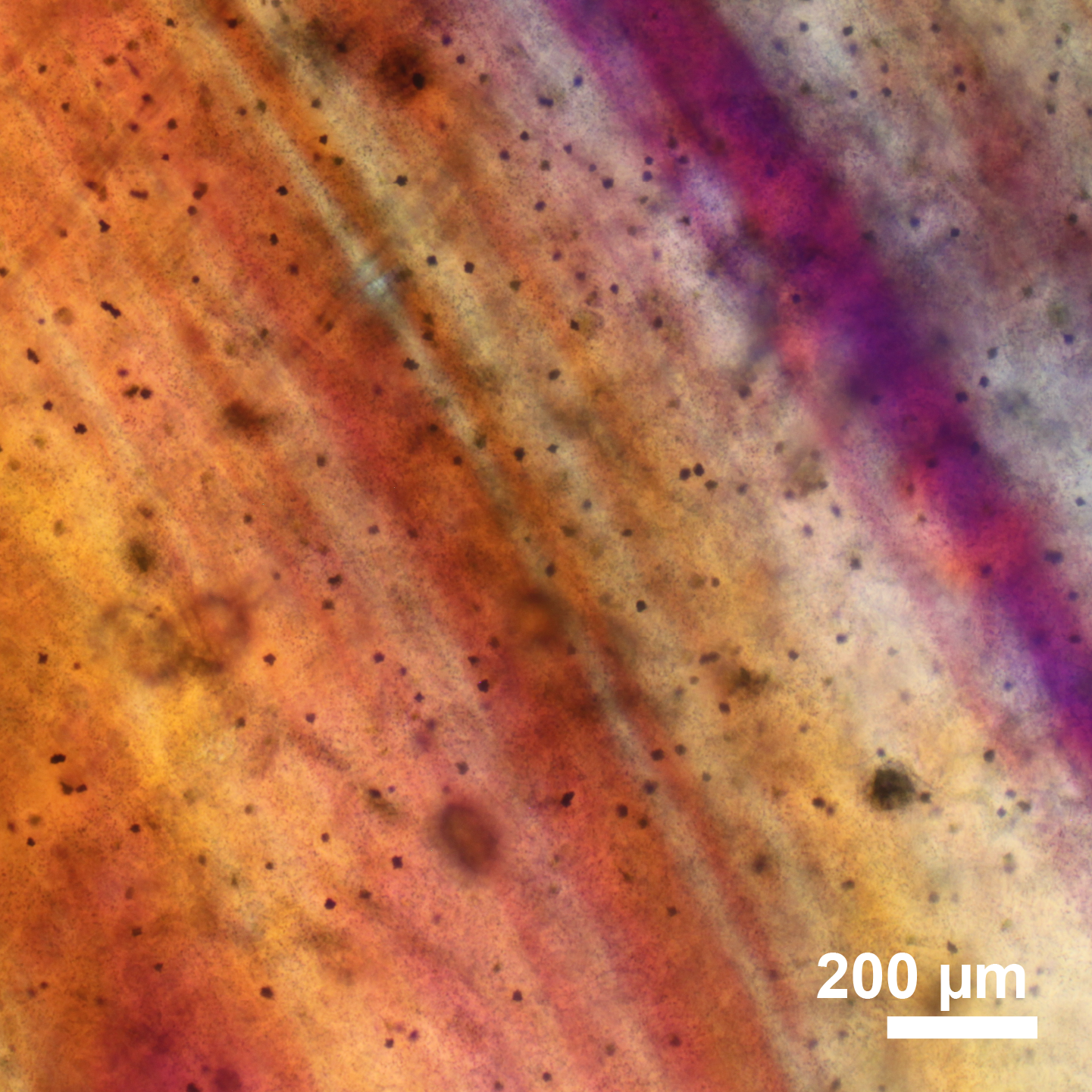
Polarized microscopy image of sea-snail egg
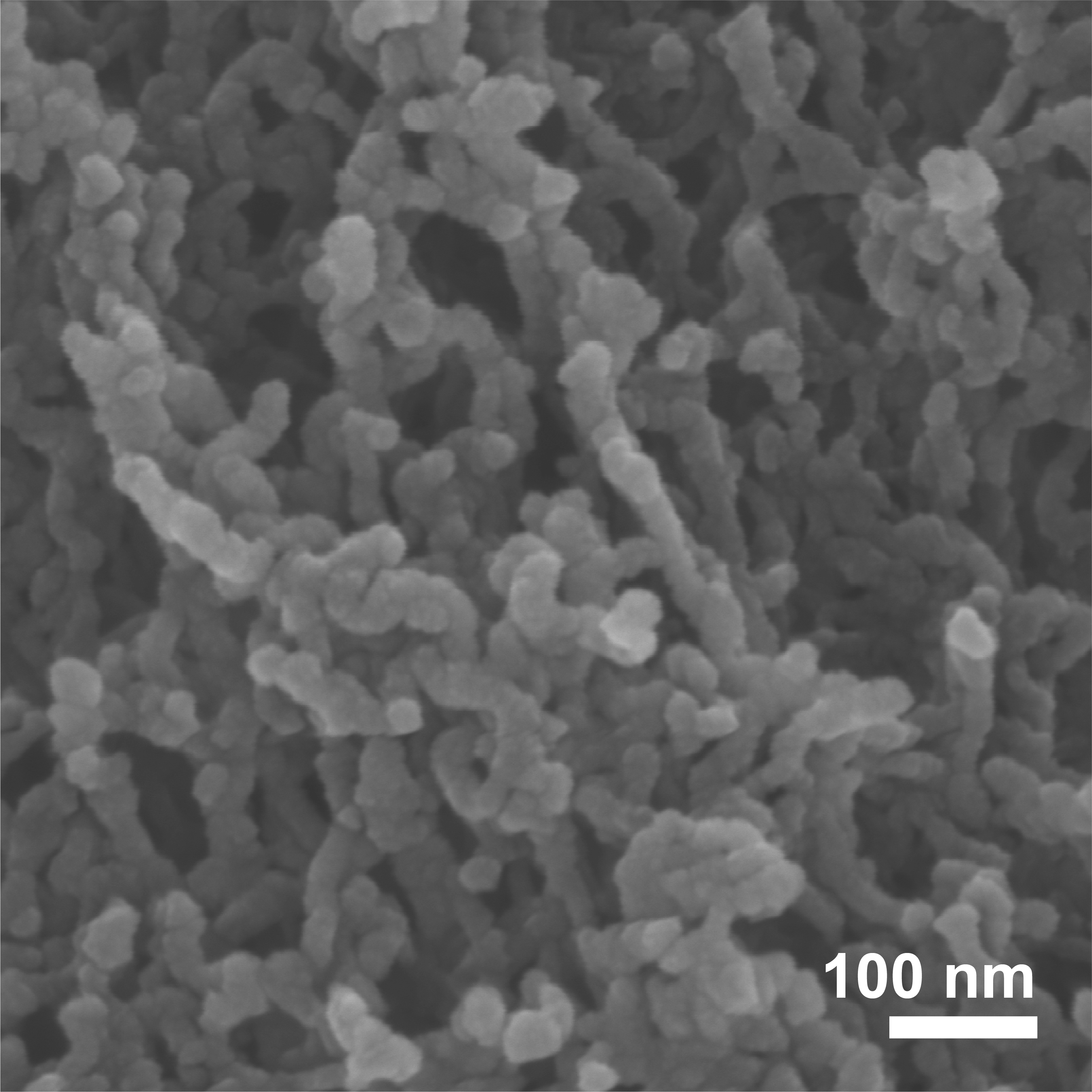
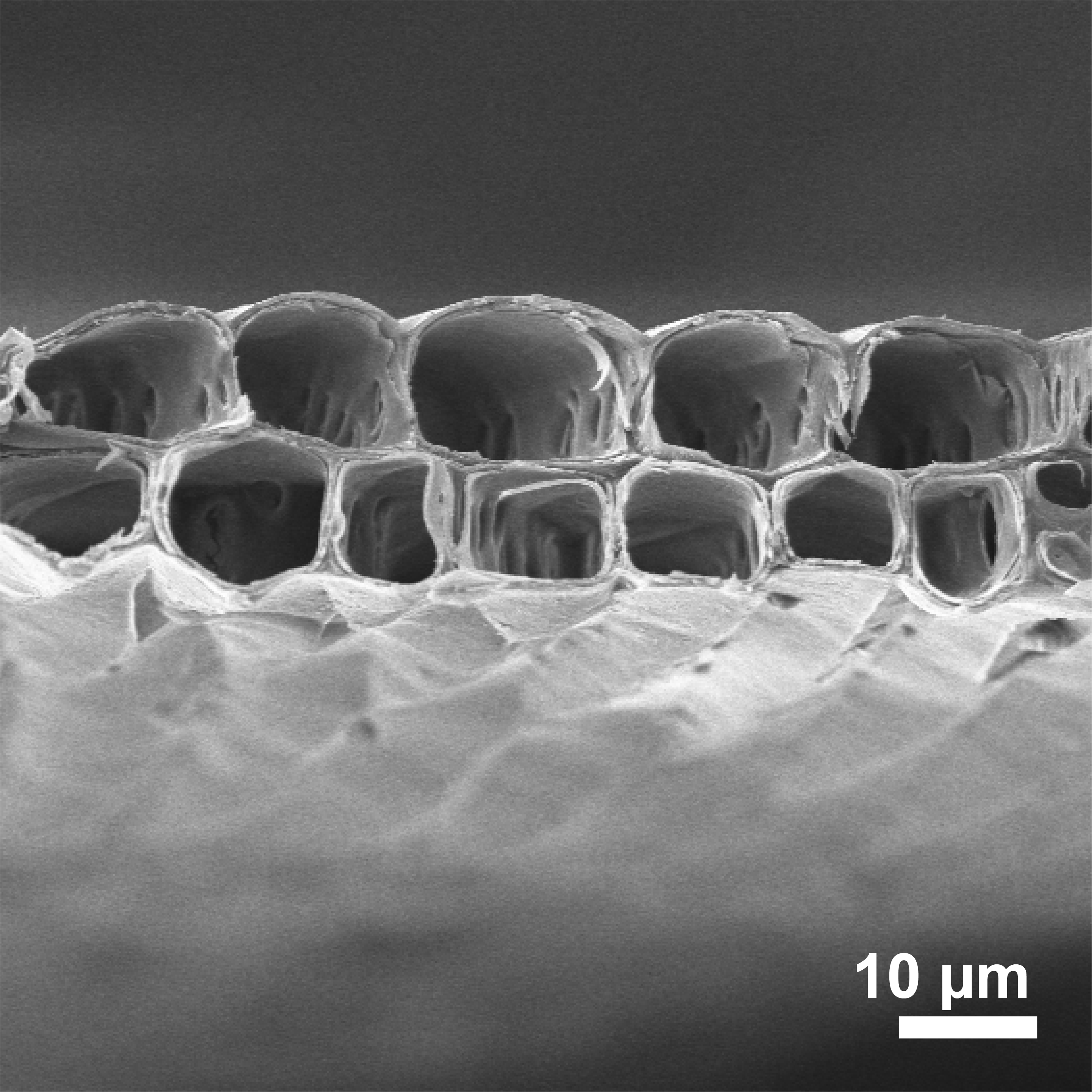
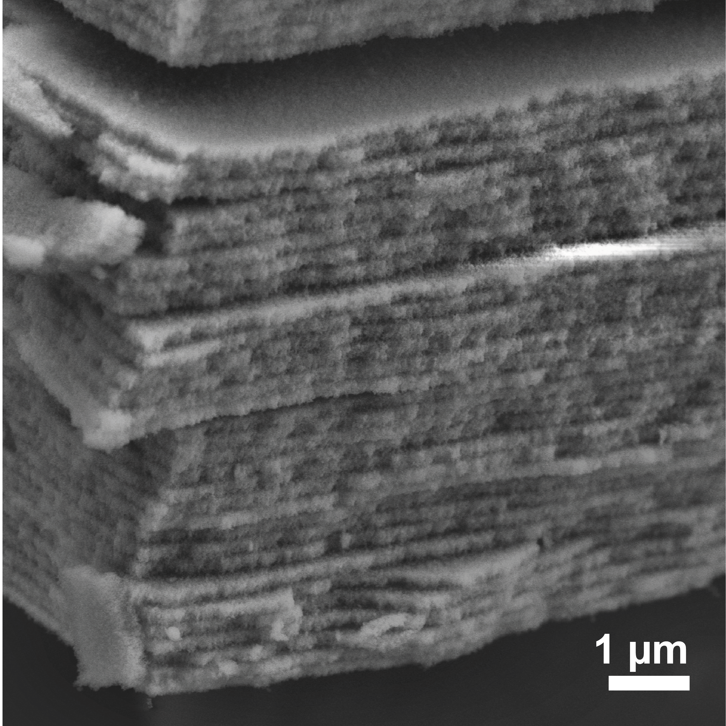
SEM image of silk-zirconia photonic crystal
Multispectral image of Lunaria annua pod
SEM image of Lunaria annua pod
Cryo-SEM image of crystals on jewel orchid
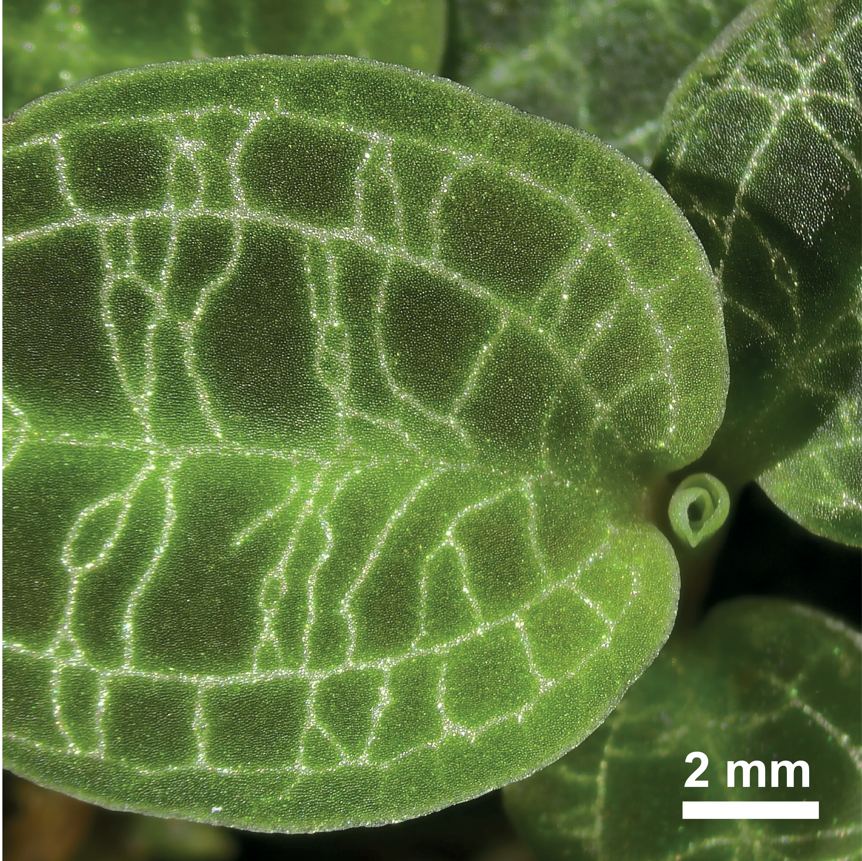
Stereomicroscopy image of Macodes petola jewel orchid
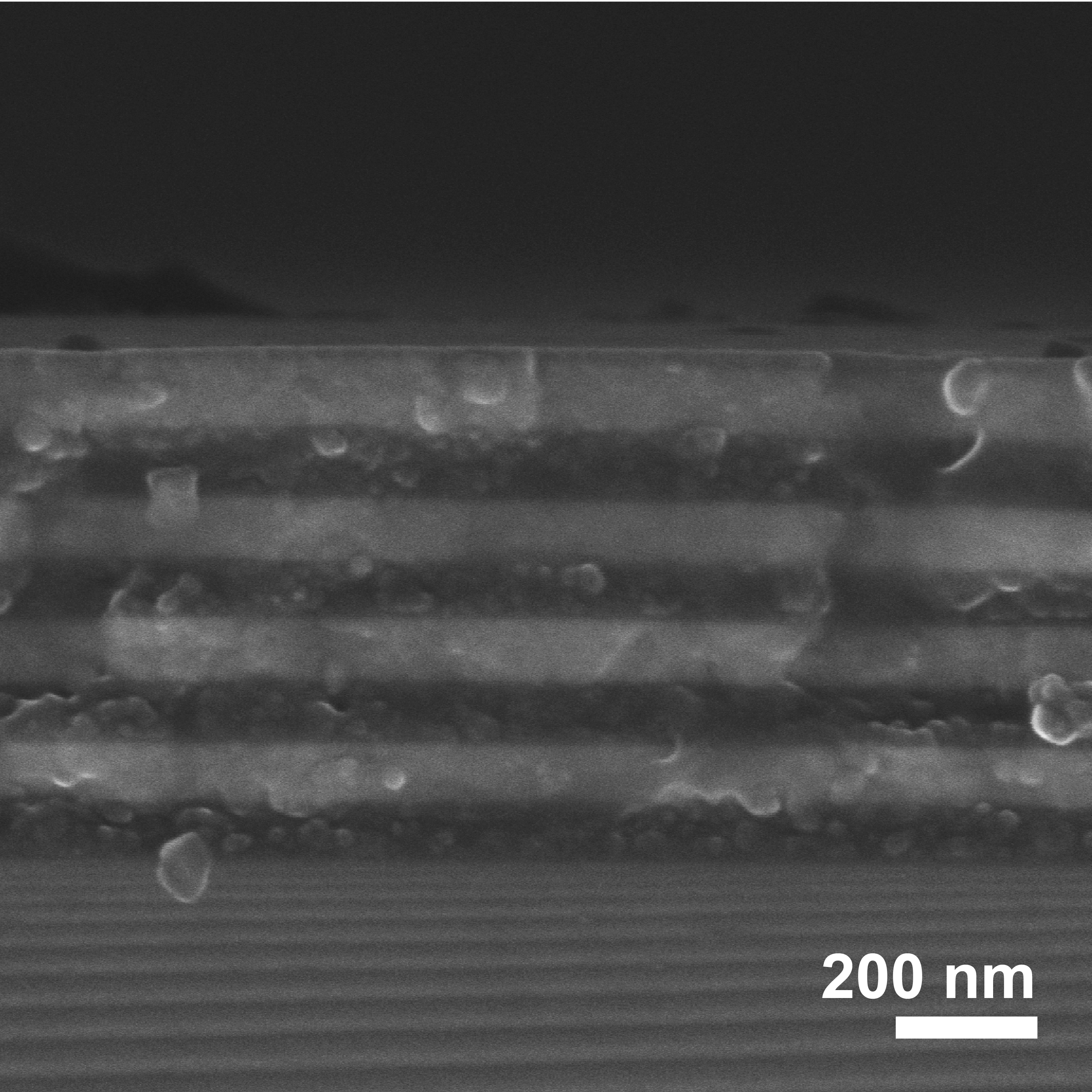
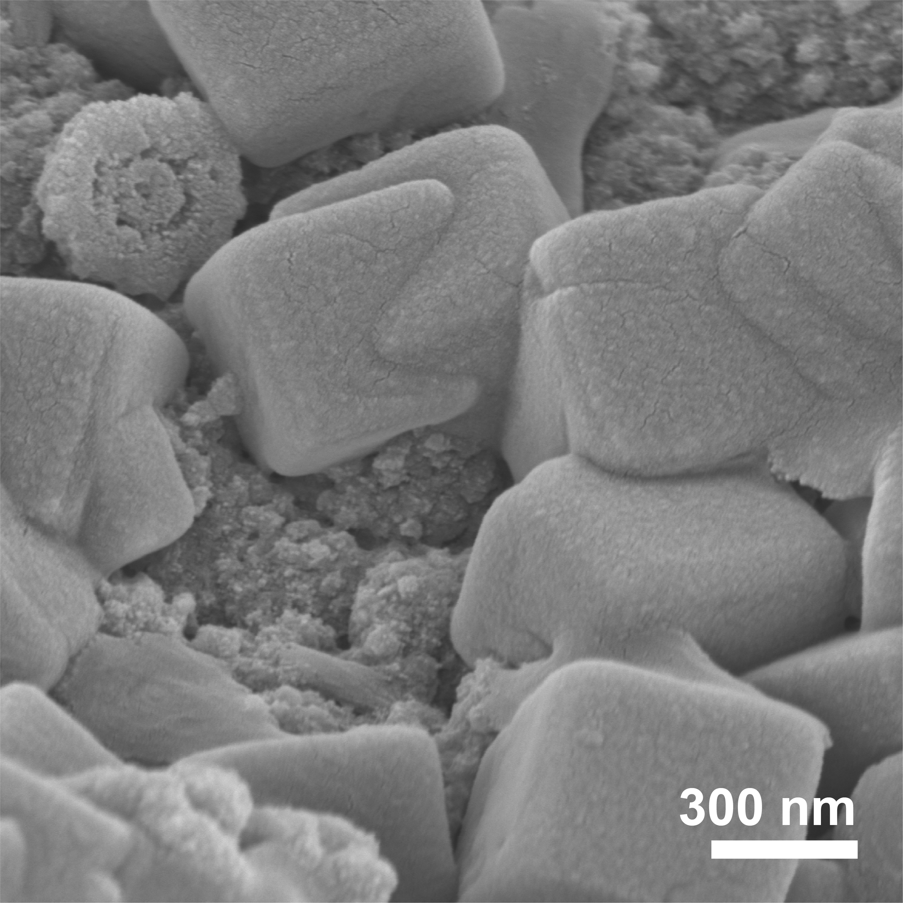
SEM image of Roman glass photonic crystal
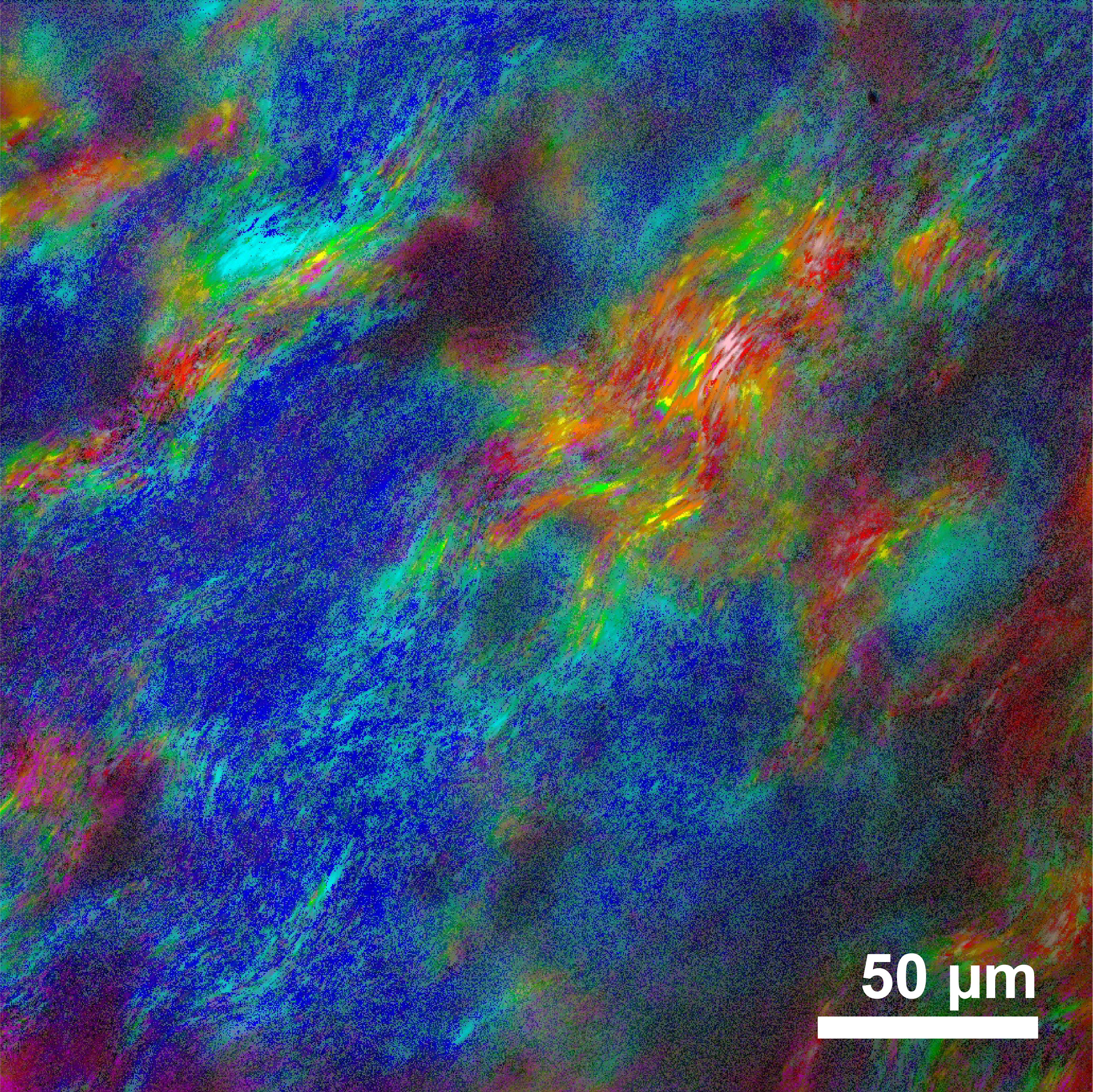
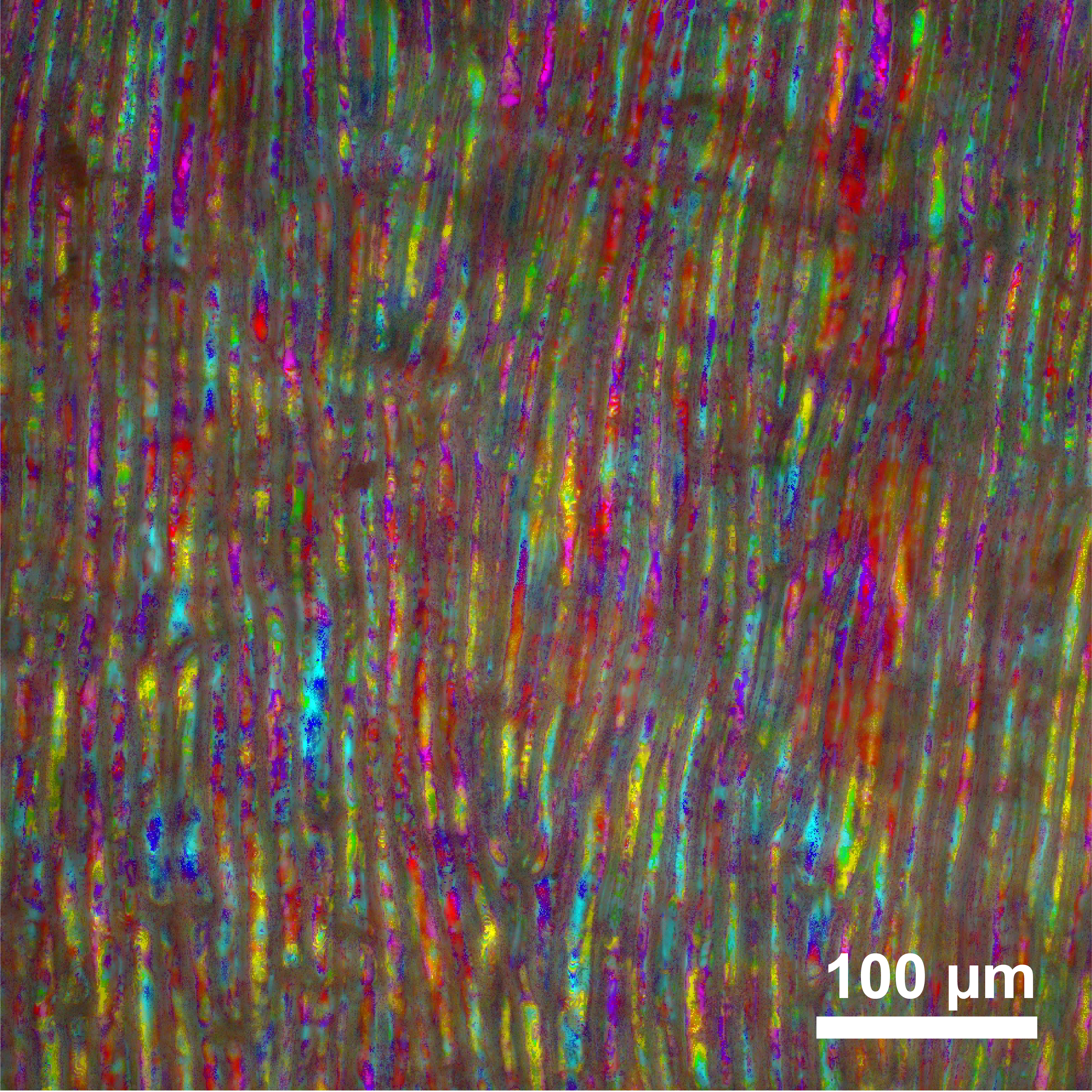
Multispectral image of Bos taurus tapetum
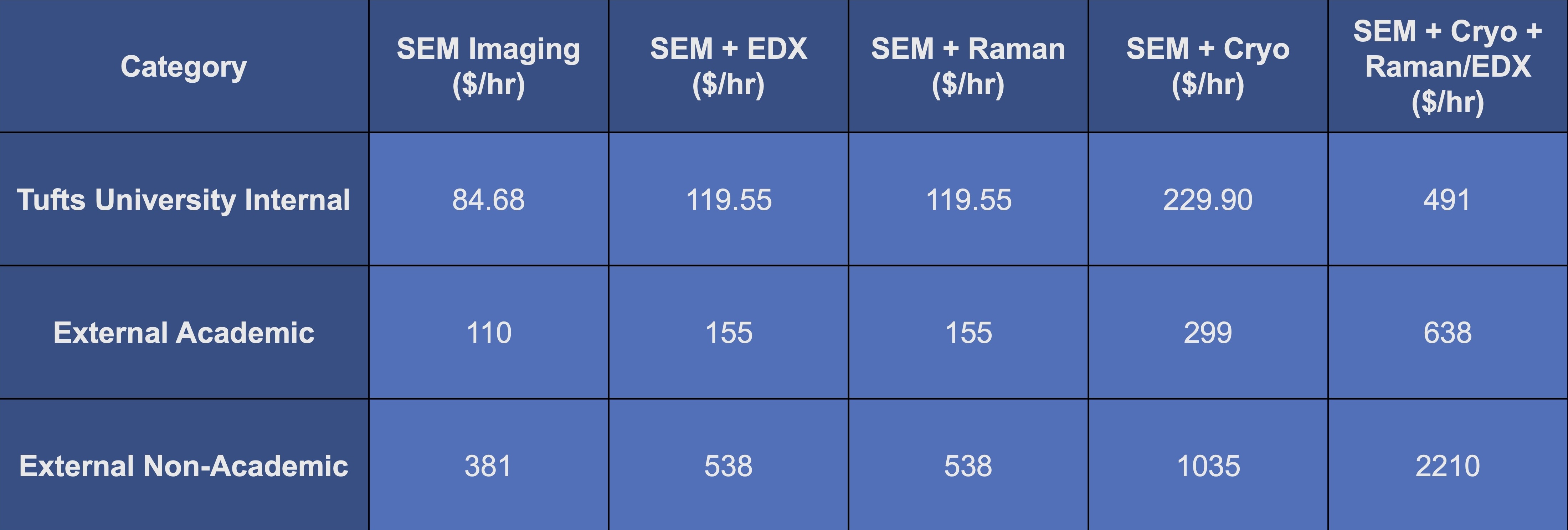
COCOON operates in assisted-use mode. All imaging sessions are carried out by trained staff who operates the microscope for you to ensure the highest quality data and optimal instrument performance.
Because sample preparation is critical to imaging outcomes, we encourage researchers to consult with us before preparing or submitting samples. Even when limited by preparation constraints, our team will obtain the best possible images and spectra.
Biosafety Level 2 (BSL-2) materials are not permitted in this facility.
For scheduling and inquiries on scientific collaborations, please contact:
Research Prof. Giulia Guidetti, giulia.guidetti@tufts.edu
Silvia Betti, silvia.betti@tufts.edu
Prof. Fiorenzo Omenetto, fiorenzo.omenetto@tufts.edu
Location
Silklab, Suite 4875
200 Boston Avenue
Medford, MA 02155